


I check the effects of adjustable parameters k0, na and
![]() on roughened images. The quantitative analysis of the effects
on the filter spectrum is provided in appendix A.
on roughened images. The quantitative analysis of the effects
on the filter spectrum is provided in appendix A.
When comparing the effects of k0 and na, I set ![]() to make
sure the filters have the same zero-frequency response. Figure
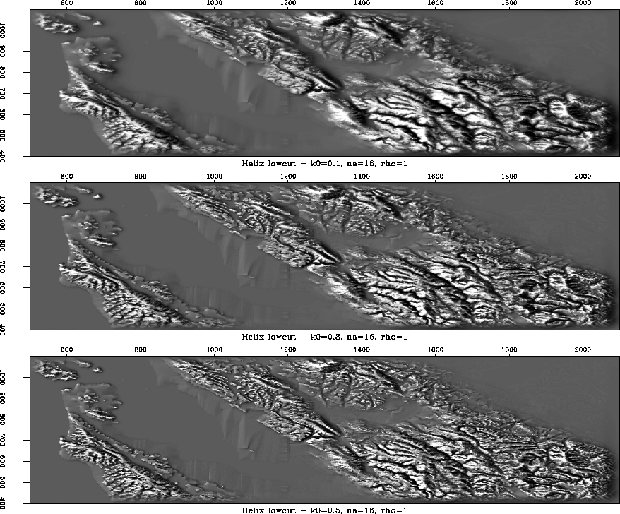
3 shows the Bay Area map created with
different k0. As k0 increases, more low-frequency components
were removed and the detailed structure turns out to be the main
focus of the map. This indicates the cut-off frequency increases as
k0 increases. In other words, k0 governs the cut-off frequency.
When k0 remains the same, filters with different na can create
very similar results if the zero-frequency response is the same by
adjusting
to make
sure the filters have the same zero-frequency response. Figure
3 shows the Bay Area map created with
different k0. As k0 increases, more low-frequency components
were removed and the detailed structure turns out to be the main
focus of the map. This indicates the cut-off frequency increases as
k0 increases. In other words, k0 governs the cut-off frequency.
When k0 remains the same, filters with different na can create
very similar results if the zero-frequency response is the same by
adjusting ![]() , as shown in the middle and bottom plots in Figure
4. As expected,
, as shown in the middle and bottom plots in Figure
4. As expected, ![]() controls the zero-frequency
response. The larger
controls the zero-frequency
response. The larger ![]() leads to higher contrast, as shown in the
top and middle plots in Figure 4.
leads to higher contrast, as shown in the
top and middle plots in Figure 4.
 |


 |
Among the three adjustable parameters, na is the least important one
because the long filter can be replaced with a short one by adjusting ![]() .k0 controls the cut-off frequency and
.k0 controls the cut-off frequency and ![]() controls the zero-frequency
response. It is hard to tell which one is more important if I use only
this information. From the quantitative analysis, I know k0 affects
zero-frequency response significantly, but
controls the zero-frequency
response. It is hard to tell which one is more important if I use only
this information. From the quantitative analysis, I know k0 affects
zero-frequency response significantly, but ![]() does not have such
an influence on cut-off frequency. So k0 is the most important
parameter.
does not have such
an influence on cut-off frequency. So k0 is the most important
parameter.
For the enhanced helix low-cut filter, it is very reasonable to choose
parameter k0 first, then ![]() and na.
and na.
When roughening the image with the helix low-cut filter, the key point is to choose the cut-off frequency f0 or the adjustable parameter k0. My suggestion is that if the lowest frequency component to be preserved is fL, k0 should be
| |
(5) |
Since the short filter is made equivalent to the long filter by adjusting
![]() , I suggest the use of
, I suggest the use of ![]() based on the balance of
computation costs and symmetric features.
based on the balance of
computation costs and symmetric features.
With k0 and na determined, I can find ![]() according to Equation
(13)
according to Equation
(13)
| |
(6) |
Figure 5 consists of three maps of the Bay Area.
The top portion is a topographic map of the Bay Area. The bottom plot
is the preferred result. From one slice of the Bay Area topographic map,
I know one main low frequency component is about 0.4. So I chose
k0 = 0.3 to remove the lower frequency. I chose na = 16. In order
to obtain the highest contrast, I chose ![]() . The middle one is the
reference plot with k0 = 0.1, na =16 and
. The middle one is the
reference plot with k0 = 0.1, na =16 and ![]() . I notice that
the bottom plot removes more low frequency components than the middle
one and has clearer details, as predicted by the theory.
. I notice that
the bottom plot removes more low frequency components than the middle
one and has clearer details, as predicted by the theory.
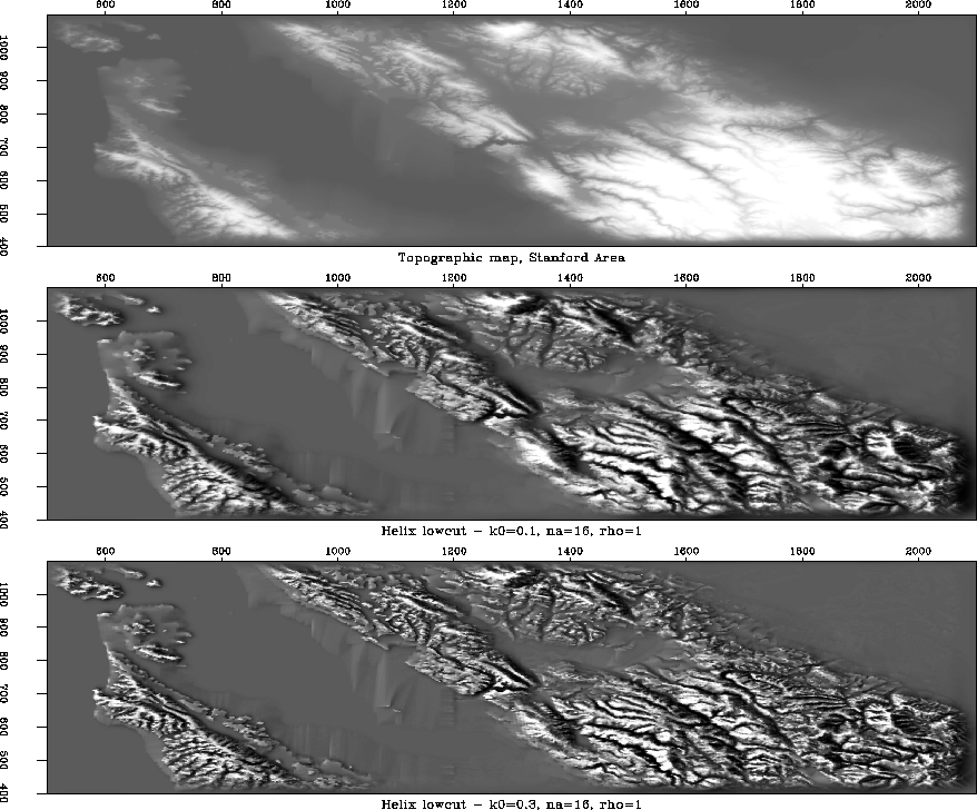 |
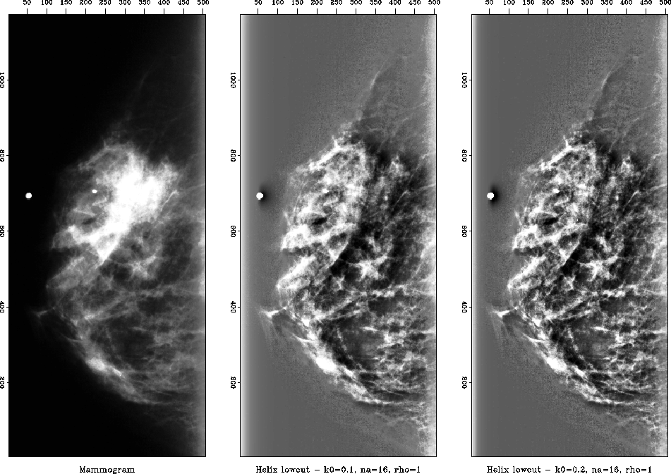
Figure 6 consists of a normal mammogram and the
roughened images.
The main low frequency component of the mammogram slice is about
0.3, so I choose k0 = 0.2 in the right plot as the preferred
result, and use k0 = 0.1 in the middle as reference.
I use ![]() to achieve the highest contrast.
to achieve the highest contrast.
 |